Understanding How InView Imaging Turns Images Into Answers
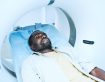
When you come to InView Imaging for an MRI, CT, ultrasound, X-ray, DEXA, or PET/CT exam, your visit doesn’t end when you walk out the door. In many ways, the most important part of your imaging exam happens after the scan—when a radiologist interprets your images and turns them into a clear, clinically useful report for your doctor.
Patients often wonder:
- Who reads my scan?
- What exactly does the radiologist look for?
- How do they know what’s normal and what isn’t?
- When do I get my results?
- What does the report actually include?
This guide explains what happens behind the scenes at InView Imaging after your scan is completed—and how our radiologists work to ensure your doctor receives accurate, timely, and meaningful results.
- Your Images Are Reviewed by a Fellowship-Trained Radiologist
Radiologists are medical doctors who specialize in diagnosing disease and injury using imaging exams. At InView Imaging, your images are reviewed by board-certified, fellowship-trained radiologists with expertise across subspecialties, including:
- Neuroradiology (brain, spine, nerves)
- Musculoskeletal imaging (joints, ligaments, tendons, sports injuries)
- Body imaging (abdomen, pelvis, organs)
- Chest imaging (lungs, heart, thorax)
- Oncology imaging (tumors, staging, surveillance, PET/CT)
- Women’s imaging (breast and pelvic conditions)
This subspecialty structure allows InView to match your exam with the radiologist best trained to interpret that type of study—leading to more accurate, clinically helpful results.
For example:
- A knee MRI goes to an MSK radiologist.
- A brain MRI goes to a neuroradiologist.
- A PET/CT exam (InView San Jose) goes to a radiologist trained in oncology imaging.
This targeted expertise is a key benefit of choosing a community imaging center with deep physician involvement and specialized training.
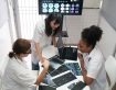
- High-Resolution Images Are Carefully Reviewed
Once your scan is completed, the images are transmitted securely to our radiology reading workstations. These are advanced diagnostic systems with high-resolution monitors designed specifically for medical imaging interpretation.
A radiologist will review your exam by:
- Examining each image and sequence in detail
- Comparing your current images to any prior scans you may have had
- Looking at multiple imaging planes and sequences
- Evaluating both the area of concern and surrounding structures
- Considering your symptoms and clinical history
This process is meticulous. A single MRI or CT scan can include hundreds or even thousands of images. Radiologists evaluate every one to look for patterns, abnormalities, or changes that may explain your symptoms.
- Correlating Clinical Information With Imaging Findings
A radiologist doesn’t just “look at pictures.” They combine:
- Your symptoms
- The reason for the exam
- Any clinical notes from your provider
- Your prior imaging results
- Your medical history (when available)
This context is essential. The same imaging finding may have different meanings depending on your symptoms or clinical scenario.
For example:
- A disc bulge on an MRI may be normalin someone without symptoms.
- The same disc bulge could explain sciatica in someone with leg pain.
Radiologists at InView aim to produce reports that align with your doctor’s question and help guide next steps in your care.
- Inside the Radiologist’s Report: What It Actually Contains
Most patients never see the actual radiology report, but understanding its structure can help you feel more informed and empowered.
A typical report from InView Imaging includes the following sections:
- Examination Details
This describes:
- The type of study performed
- Whether contrast was used
- The technique and sequences used
- The date of the exam
Example:
MRI Lumbar Spine Without Contrast, using T1, T2, STIR, and axial sequences
- Clinical Indication
This tells the radiologist what your doctor is looking for, such as:
- “Low back pain radiating to left leg”
- “Chronic knee pain, rule out meniscal tear”
- “Follow-up of known lung nodule”
- “Headaches with visual changes”
This helps focus the interpretation on what matters most.
- Findings Section
This is the most detailed part of the report. The radiologist describes:
- Normal anatomical structures
- Any abnormalities
- Measurements
- Levels, segments, or regions involved
- Additional observations
For example, in a knee MRI the findings might detail:
- Menisci
- ACL/PCL
- Cartilage surfaces
- Bone marrow signal
- Surrounding soft tissues
In a CT abdomen/pelvis, the radiologist may describe:
- Liver, gallbladder, spleen
- Kidneys, bowel, bladder
- Lymph nodes and vessels
- Bones and soft tissues
This section is written in technical language for your doctor.
- Impression (Summary)
This is the part most physicians—and many patients—read first.
It gives:
- A clear summary of major findings
- A diagnosis, when appropriate
- A differential diagnosis when more than one condition is possible
- Recommendations for follow-up
- Urgent findings highlighted clearly
Example:
Impression: Findings consistent with a medial meniscal tear. Mild osteoarthritis. No ligament injury.
This section is designed to be concise and actionable.
- When Do Results Arrive?
At InView Imaging, our goal is to provide efficient, timely results to your referring provider, typically within 24–48 hours for most outpatient studies. More urgent exams can be read sooner depending on the clinical situation.
Your physician receives:
- The full radiology report
- Access to images
- The ability to consult with our radiologist directly
Your doctor will then review the results with you—explaining what they mean, how they relate to your symptoms, and what the next steps might be.
- How Radiologists Collaborate With Referring Physicians
InView Imaging fosters strong communication with referring providers. When needed, our radiologists:
- Call physicians directly about urgent findings
- Discuss complex cases
- Recommend additional imaging if helpful
- Clarify subtle or ambiguous findings
- Support clinical decision-making
This collaboration ensures your imaging results are interpreted in context—not in isolation.
Our radiologists view themselves as partners in your care, working with your doctor to give you the clearest possible picture of your health.
- What If Something Unexpected Is Found?
Not all imaging findings match the reason for your exam. Sometimes radiologists discover incidental findings—abnormalities unrelated to your symptoms.
Examples include:
- Small thyroid nodules
- Benign liver cysts
- Kidney stones
- Mild degenerative changes
Most incidental findings are benign, but some require follow-up.
At InView Imaging:
- These findings are clearly noted in the report
- Recommendations are made when appropriate
- Your doctor determines whether follow-up imaging or evaluation is needed
We aim to balance clarity with clinical relevance—identifying what matters while avoiding unnecessary alarm.
- Seeing Your Images
Many patients like to view their own images, and InView Imaging makes this easy:
- You may request digital access or a copy of your imaging
- Physicians can share key images during follow-up visits
- Our staff can help you understand how to access your records
Seeing your images can be interesting, reassuring, and empowering.
- Why the Radiologist’s Role Matters
Although patients rarely meet their radiologist face-to-face, the radiologist plays a critical role in your diagnosis and care plan.
Radiologists at InView Imaging:
- Provide highly specialized interpretation
- Ensure accuracy and clarity
- Support early diagnosis of serious conditions
- Help avoid unnecessary procedures
- Contribute to better treatment decisions
Behind every image is a highly trained physician analyzing hundreds of details so your doctor can provide the best care possible.
- The InView Imaging Difference
Patients and physicians tell us that what sets InView Imaging apart is our:
- Subspecialty-trained radiologists
- Efficient, patient-friendly workflow
- Quick, easy scheduling
- Modern MRI options (open MRI in Concord, wide-bore MRI in San Ramon)
- PET/CT in San Jose for oncology evaluations
- Clear, actionable radiology reports
- Local, physician-owned, community-focused care
Every scan is treated with the same level of attention and expertise we would want for our own families.
Final Thoughts: Turning Images Into Answers
Your imaging exam is only the first step. The real clarity comes from the radiology report—created by experienced physicians who interpret the images with care, precision, and clinical insight.
At InView Imaging, we take pride in delivering reports that help:
- Diagnose problems earlier
- Guide treatment
- Support specialists
- Give patients confidence in their careIf you ever have questions about your imaging or your experience, our team is here to support you.